Дирофиляриоз оболочек яичка под маской синдрома отечной и гиперемированной мошонки
Кафедра детской хирургии Российской медицинской академии последипломного образования, Тушинская детская городская больница, Москва, Россия.
Дирофиляриозом болеют собаки, кошки, дикие плотоядные и иногда человек. Возбудители заболевания - нематоды семейства Filariidae: Dirofilaria immitis, Dirofilaria repens и другие. D. repens является облигатным гельминтом собак, паразитирующим в подкожной соединительной ткани. Промежуточные хозяева - комары Aedes aegypti и Anopheles maculipennis. Заболевание регистрируют чаще в районах с теплым и влажным климатом [1].
В литературе встречаются описания редких наблюдений поражения органов мошонки глистной инвазией [2]. В нашем исследовании было 2 случая обнаружения гельминтов оболочек яичка во время операции ревизии органов мошонки. При паразитологическом исследовании гельминта определена дирофилярия. Описание этих случаев представляет интерес как казуистическое заболевание, нехарактерное для центрального региона России.
Больной С., 11 лет, поступил в клинику 14.09.01. с жалобами на покраснение, припухлость, болезненность в правой половине мошонки в течение 2-х дней. При поступлении состояние не нарушено. Местно: умеренное увеличение в объеме правой половины мошонки, отечность, гиперемия кожи, при пальпации выраженный плотный отек оболочек, преимущественно по передней поверхности. Пальпировалось возрастных размеров яичко, подвижное, безболезненное, умеренная болезненность в области верхнего полюса. Выполнено УЗИ органов мошонки, которое выявило увеличение правого придатка в области тела и хвоста, усиление кровотока; значительное утолщение оболочек яичка справа, под оболочками - умеренное количество свободной жидкости с дисперсной взвесью. Достоверно дополнительных образований не определялось.
При оперативной ревизии правой половины мошонки в оболочках яичка обнаружен гельминт размерами 10,0 см - 0,2 см, белого цвета, подвижный. При ревизии органов мошонки патологии не выявлено, удалена неизмененная гидатида. Течение послеоперационного периода гладкое. Проводилась противовоспалительная терапия. Послеоперационная рана зажила первичным натяжением. Выписан на 6 сутки в удовлетворительном состоянии. Паразитологическое исследование: дирофилярия. Катамнез через 1 месяц с УЗИ органов мошонки не выявил патологических изменений.
Больной С., 9 лет, 24.03.99. с жалобами на покраснение, припухлость левой половины мошонки обратился в приемное отделение клиники через 5 часов от начала заболевания. В анамнезе 25.02.99. операция - удаление перекрученной гидатиды левого яичка. Состояние при поступлении не нарушено. Местные изменения характеризовались гиперемией кожи левой половины мошонки, в области левого яичка пальпировалось опухолевидное образование плотной консистенции, безболезненное. При ультразвуковом исследовании 25.03.99. между верхним полюсом яичка и головкой придатка определялось эхоплотное включение размером 2 мм на фоне свободной жидкости. В оболочках левого яичка определялось дополнительное объемное образование овальной формы, размерами 26 мм - 13 мм, средней эхогенности, с эхоплотными линейными включениями по центру. 26.03.99. произведена оперативная ревизия левой половины мошонки. По послеоперационному рубцу вскрыты оболочки яичка, удалены лигатура оболочек и лигатура на верхнем полюсе яичка. При пальпации определялся инфильтрат выше раны. При его ревизии обнаружен подвижный гельминт длиной около 5 см. Произведено иссечение измененных тканей, ушивание раны. Течение послеоперационного периода гладкое. Проводилась противовоспалительная терапия, получал аспирин, супрастин. Выписан на 7 сутки в удовлетворительном состоянии. При катамнестическом исследовании через 2 года жалоб, местных патологических изменений не выявлено, что подтверждено данными УЗИ органов мошонки.
Мы приводим описания случаев дирофиляриоза оболочек яичка с целью продемонстрировать их проявления, характерные для синдрома отечной и гиперемированной мошонки у мальчиков. При дифференцировке заболеваний, представленных отеком и гиперемией половины мошонки, необходимо помнить о возможности этой патологии. В диагностике дополнительную информацию предоставляет ультразвуковое исследование в В-режиме. Эксплорация является основным диагностическим и лечебным приемом. Дополнительные терапевтические меры при этом заболевании не показаны.
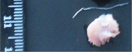
(Фото Стоногин С.В.) Взрослый самец Dirofilaria repens (вверху) и фрагмент оболочек яичка (внизу).
Литература.
1. Паразитология человека. Под редакцией проф. Первомайского Г.С. и проф. Подоляна В.Я. - М.: Медицина. - 1974. - С. 301.
2. Dissanaike AS; Abeyewickreme W; Wijesundera MD; Weerasooriya MV; Ismail MM "Human dirofilariasis caused by Dirofilaria (Nochtiella) repens in Sri Lanka." Dissanaike AS - Parassitologia - 01-Dec-1997; 39(4): 375-82.
3. Logar J; Novsak V; Rakovec S; Stanisa O "Subcutaneous infection caused by Dirofilaria repens imported to Slovenia." Logar J - J Infect - 01-Jan-2001; 42(1): 72-4.
4. Mussner W., Bosch J., Buhl D., Neuweiter J., Bandhauer K. "Filarien: eine Tropenkrankheit als Ursache fur das acute Scrotum" Urologe A. - 1997. - Vol. 36. - P. 84-86.
5. Pampiglione S; Rivasi F; Angeli G; Boldorini R; Incensati RM; Pastormerlo M; Pavesi M; Ramponi A "Dirofilariasis due to Dirofilaria repens in Italy, an emergent zoonosis: report of 60 new cases." Pampiglione S - Histopathology - 01-Apr-2001; 38(4): 344-54.
Данные об авторах:
1. Марина Витальевна Григорьева - научный сотрудник НИИ НДХиТ. grigorievamv@mail.ru, http://grigorievamv.narod.ru/ Москва, Большая Полянка 20, НИИ НДХиТ, отделение чистой хирургии.
2. Евгений Викторович Дворовенко - заведующий отделением экстренной хирургии Тушинской детской городской больницы г. Москвы, хирург высшей категории.
Адрес: 123480, Москва, Героев Панфиловцев 28, отделение экстренной хирургии. Тел.: 949 03 04 раб.
3. Стоногин Сергей Васильевич – врач-хирург хирургического отделения Тушинской детской городской больницы, кандидат медицинских наук. E-mail: svas70@mail.ru
Адрес: 125480 Москва, улица Героев Панфиловцев, дом 28, отделение хирургии. https://orcid.org/0000-0003-3531-5849.
Diagnosis and Treatment of Dirofilariasis of the Testicular Membranes in Children
M.V. Grigorieva, E.V. Dvorovenko, S.V. Stonogin, T.R. Lavrova, V.G. Supryaga, A.Yu. Komlev
Tushino Children's City Hospital, Department of Pediatric Surgery, Russian Medical Academy of Postgraduate Education
(Head of Department, Prof. V.E. Shchitinin),
Institute of Medical Parasitology and Tropical Medicine named after E.I. Marcinovsky, Moscow, Russia.
Dirofilariasis is a zoonotic, vector-borne helminthiasis of domestic and wild carnivores. Humans serve as incidental hosts, with the parasite developing to a single sexually mature stage within them (1).
The causative agent of the disease is nematodes of the Filariidae family, specifically Dirofilaria repens. D. repens is an obligate helminth of dogs, parasitizing in the subcutaneous connective tissue. Intermediate hosts are mosquitoes of the Aedes, Culex, and Anopheles genera. The disease is more common in regions with warm and humid climates. Until the mid-20th century, only a few dozen human cases of Dirofilaria repens infection were recorded. Over the past 50 years, the number of such cases has increased significantly, with the highest incidence in the Mediterranean region and Sri Lanka. In Russia, human dirofilariasis primarily occurs in the southern and southeastern regions. In the Moscow region, 11 cases were recorded between 2000 and 2002, in patients aged 4 to 72 years, including 6 males and 5 females. A relatively high frequency of D. repens localization in the male reproductive organs was identified among patients in the Moscow region (2).
Since dirofilariasis is a sporadic disease with nonspecific clinical symptoms, diagnosis is based on the morphological examination of the surgically removed helminth, posing challenges for clinical diagnosis (3). Parasitological diagnosis is difficult because microfilariae are absent in human blood. Eosinophilia is not characteristic, making laboratory diagnostics ineffective (1). Our experience in treating three patients with scrotal dirofilariasis is of interest regarding preoperative diagnosis and surgical treatment features.
Between 1999 and 2003, three patients aged 2 years 8 months, 9 years, and 11 years were hospitalized in the emergency surgery department of Tushino Children's City Hospital with the diagnosis of dirofilariasis of the testicular membranes. Upon admission, patients complained of swelling and hyperemia of one half of the scrotum for several days. With a preliminary diagnosis of scrotal edema and hyperemia syndrome, they were hospitalized in the emergency surgery department (4).
Clinical Examination and Imaging
Clinical examination revealed specific local changes in the scrotum. Palpation identified a firm, mildly tender infiltrate in the testicular membranes (scrotal wall), ranging from 5 mm to 25 mm in size, mobile, and not connected to the testis or epididymis. Notably, two patients underwent repeat surgery after initial procedures performed 2 weeks to 1 month before readmission. One underwent revision of the scrotal organs with removal of a twisted hydatid, while the other had diagnostic scrototomy with no pathological changes found in the testis or epididymis, nor was a hydatid detected. Upon readmission, the infiltrate was separate from the postoperative scar, located above and proximally to it.
Ultrasound Findings
Ultrasound examination (US) of the scrotal organs revealed specific pathological changes in two patients. Using a linear transducer with a frequency of 7.5-13 MHz on a Voluson-750 ultrasound machine (Kretz, Austria), an additional oval-shaped mass was detected, measuring 26 mm × 13 mm in the 9-year-old and 16 mm × 5 mm in the 2-year-8-month-old, with medium echogenicity and echodense linear inclusions centrally (Figures 1 and 2).
Surgical Treatment
All patients underwent emergency surgery. Upon incision of the testicular membranes, no pathological changes in the testis or epididymis were found. Revision of the infiltrate revealed dense altered tissues with a live, thread-like white helminth at the center. The helminth was removed, and excision of the pathological tissue of the testicular membranes was performed (Figure 3).
Postoperative recovery was uneventful for all patients. Scrotal swelling subsided by days 3-5 with treatment (aspirin, diazolin, physiotherapy). Surgical wounds healed satisfactorily, and patients were discharged in good condition on postoperative days 6-7.
Parasitological examination confirmed Dirofilaria repens in all three cases: two female worms and one male worm were identified. Histopathological examination of the affected testicular membranes revealed inflammatory infiltrates dominated by eosinophils and lymphocytes.
Discussion
Acute testicular diseases presenting with swelling, hyperemia, and pain in the scrotum are common in children and often require emergency surgical intervention. Differential diagnosis includes torsion of a testicular hydatid, epididymitis, epididymo-orchitis, testicular torsion, and traumatic injuries.
Among acute testicular conditions in children, torsion of a testicular hydatid or epididymis was the most common pathology in our study (57%). Hydatids are embryological remnants of the urogenital system, including the appendix testis (Morgagni hydatid), appendix epididymis, paradidymis (Giraldes’ organ), and aberrant ductules (5). Epididymitis and epididymo-orchitis accounted for 18% of cases, testicular torsion for 16%, and scrotal trauma for 9%.
Ultrasound is a highly informative, non-invasive, and safe imaging method for diagnosing acute testicular conditions. In our study, ultrasound criteria were established based on examinations of 219 children with acute testicular diseases.
Conclusions
-
Dirofilariasis should be considered in the differential diagnosis of acute testicular diseases in children.
-
Clinical features of testicular dirofilariasis in children include progressive scrotal swelling and hyperemia, absence of pain, normal body temperature, and unremarkable laboratory tests. The key clinical manifestation is a small, painless, localized scrotal wall infiltrate, independent of the testis, epididymis, or spermatic cord.
-
Preoperative diagnosis of testicular dirofilariasis is possible via ultrasound, which reveals an additional oval or spindle-shaped heterogeneous mass (5-30 mm), with echodense linear inclusions centrally and no blood flow on color Doppler imaging.
-
Surgical exploration for suspected testicular dirofilariasis should include careful revision of the infiltrate, as the helminth is not attached to the testis or epididymis.